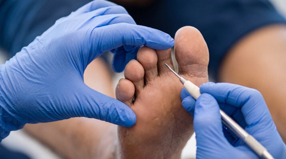
A diabetic foot ulcer appointment involves more than cleaning the wound and applying a dressing. You will learn how clinicians assess infection, blood flow, nerve damage and pressure, how they choose treatment, and which changes require urgent medical attention.
Key takeaways
- The first visit assesses urgency, infection, circulation and barriers to healing.
- Clinicians examine the ulcer, surrounding skin, pulses, sensation and the rest of the foot.
- Infection findings and circulation problems determine which tests and treatments you need.
- Follow-up checks wound size, tissue, drainage, pain and healing progress.
What happens during the first diabetic ulcer treatment consultation?
The first appointment is a triage assessment, not simply a dressing change. Before examination begins, the clinician takes a history to judge urgency, infection risk, circulation and factors delaying healing.
1. Expect questions about when the ulcer began, whether it is enlarging, and whether you have pain, drainage, odour, fever, chills or increasing difficulty walking.
They will ask about glucose readings or HbA1c, prescribed medicines, smoking, kidney disease, previous ulcers or amputations, footwear, daily walking, work demands, balance and treatments already tried, including dressings, antibiotics, debridement or home remedies.
2. The clinician checks for rapidly spreading redness, severe or new pain, black or grey tissue, heavy drainage, fever, confusion, vomiting or feeling acutely unwell. Suspected sepsis, a deep abscess, gangrene, severe loss of blood flow or deep bone infection requires same-day hospital or specialist assessment, not routine follow-up.
3. Bring medication details, glucose records, previous reports, the shoes you use and the name of every antibiotic or dressing applied. The clinician then examines the wound and foot, measures the ulcer and decides whether tests, urgent treatment or off-loading are needed.
What happens during diabetic foot ulcer treatment depends on these findings, so do not cover the ulcer with a new product immediately beforehand. A diabetic ulcer treatment consultation may lead directly to hospital referral when warning signs are present.
How is the ulcer and the rest of the foot examined?
The examination follows a set order: the clinician documents the wound, inspects the entire foot, tests sensation, then assesses blood flow.
1. They measure the ulcer’s length, width and depth and record its location. They inspect the edges, callus, deformity and surrounding skin, looking for exposed tendon or bone, slough, dead tissue, drainage, odour, gangrene, swelling, warmth, redness, colour change and skin breakdown elsewhere.
A probe-to-bone assessment may be performed when the ulcer is deep or bone infection is suspected.
2. They test protective sensation by pressing a 10-g monofilament against specific areas of the sole and toes. You may also have vibration, reflex or temperature checks when needed; numbness means pressure or injury can continue without warning.
3. They assess circulation by checking foot pulses, skin temperature and colour. Palpable pulses alone do not reliably exclude peripheral artery disease, so the clinician may order an ankle-brachial index, Doppler waveforms, toe pressure or toe-brachial index. Diabetes-related arterial calcification can make an ankle-brachial index falsely high.
4. These findings shape the diabetic foot ulcer treatment process. An ulcer exposed to repeated pressure, a numb foot or poor perfusion needs off-loading, closer monitoring or vascular review, while a shallow wound with intact sensation and circulation follows a different plan.
How do clinicians identify infection and decide which tests are needed?
What happens during diabetic foot ulcer treatment depends first on whether infection is present: a clean ulcer is not automatically infected, and antibiotics do not make an uninfected ulcer heal.
Clinicians look for local swelling or induration, warmth, erythema, tenderness or pain, and purulent discharge. Neuropathy and poor circulation can blunt these signs, so a quiet-looking wound does not prove that deeper infection is absent.
A superficial swab cannot replace examination; it may grow organisms living on the surface rather than those causing deep infection. If infection is suspected, the preferred culture is tissue collected after the ulcer is cleansed and debrided.
| Investigation | Purpose | When it applies |
|---|---|---|
| Full blood count, C-reactive protein and kidney function | Assess systemic inflammation and medication safety | Suspected infection or treatment planning |
| Foot X-ray | Detect gas, a foreign body, deformity or later bone changes | Initial assessment of deeper problems |
| MRI | Assess deep tissue infection or osteomyelitis in greater detail | Bone or deep infection remains suspected |
| Vascular imaging | Map narrowed or blocked arteries | Revascularisation is being considered |
Same-day or emergency escalation is needed for:
- Suspected sepsis or necrotising infection
- Compartment syndrome or a deep abscess
- Gangrene or severe limb ischaemia
- Suspected deep bone infection
These findings can lead to hospital admission, urgent drainage or debridement, intravenous antibiotics, and vascular, orthopaedic or surgical review. The test choice follows the clinical question; no single scan replaces examination.
What are the diabetic foot wound care steps after assessment?
After assessment, the diabetic foot wound care steps follow the wound’s depth, infection status, circulation and pressure exposure. The diabetic foot ulcer treatment process commonly includes:
1. Cleanse the wound with the solution and technique prescribed. Do not scrub deep tissue or apply household antiseptics without instructions.
2. Remove dead tissue, slough and callus when appropriate. Debridement exposes the wound base and removes pressure-maintaining tissue; sharp or surgical debridement is selected according to blood flow, pain, bleeding risk, infection and clinical findings. Severe ischaemia needs assessment before aggressive debridement.
3. Apply a dressing chosen for the drainage and wound tissue. Changes may be daily or less frequent, depending on the product, exudate and care plan; no dressing is universally best. Protect surrounding skin with a prescribed barrier film or other suitable product.
4. Expect mild soreness, slight bleeding or a larger-looking wound after debridement because hidden edges and depth become visible. Contact the treating team promptly for heavy bleeding, worsening pain, spreading redness, fever, foul drainage or rapidly increasing size.
5. Relieve pressure by reducing walking and using a prescribed removable cast walker, total-contact cast, therapeutic shoe, felted foam or another off-loading device. A soft dressing does not make normal activity safe.
6. Review footwear for fit, balance and pressure points, then check the foot and device daily. A removable boot works only when you wear it.
7. Coordinate glucose management with the diabetes team; glucose control supports healing but does not replace cleansing, dressings, debridement or off-loading.
How is healing monitored after the first treatment?
Healing is monitored through active reassessment, not by waiting for the next dressing change. At each scheduled review, the clinician remeasures the ulcer’s length, width and depth, checks tissue quality and drainage, looks for infection, inspects pressure points and confirms that the off-loading device is being used safely.
The diabetic foot ulcer treatment process may include:
- A removable boot, total-contact cast or therapeutic footwear, selected for the ulcer’s location, circulation, balance and ability to use the device safely.
- Mobility aids, reduced activity and clear instructions to inspect both feet and the device every day.
- Dressing changes and pressure relief adjusted when the wound, drainage or surrounding skin changes.
Glucose management supports healing but does not replace wound care, pressure relief, infection treatment or restoration of blood flow. The team may review glucose readings, diabetes medicines, smoking, nutrition, footwear, callus prevention and balance.
If the wound does not improve, clinicians reconsider the diagnosis, pressure exposure, circulation, infection, bone involvement and adherence. Poor perfusion can lead to vascular referral for revascularisation; restoring blood flow does not replace off-loading or infection control.
When comparing a consultation with Dr Aditya Phadke, ask whether it includes documented measurements, 10-g monofilament sensation testing, circulation assessment, an off-loading plan, dressing instructions and a dated review schedule. These details reveal the care process better than a general claim about advanced treatment.
Frequently asked questions
What happens during the first diabetic ulcer treatment consultation?
The clinician takes your medical history and assesses urgency, infection risk, circulation and factors that could delay healing before treating the wound.
How is a diabetic foot ulcer examined?
The clinician examines the ulcer and surrounding skin, checks the rest of the foot, and assesses circulation and sensation.
How do clinicians decide which diabetic foot ulcer tests are needed?
They use examination findings, infection signs and circulation concerns to determine whether further tests are needed.
What are the diabetic foot wound care steps after assessment?
Care follows the assessment and can include wound cleaning, dressing, pressure management and treatment of infection or circulation problems.
How is healing monitored after the first treatment?
Follow-up appointments compare wound size, tissue, drainage, pain and other findings to assess healing and adjust care.
Keywords